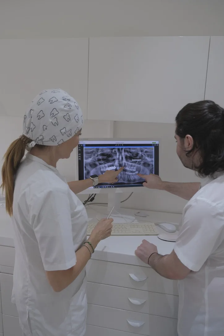

2D Digital Panoramic & 3D CBCT Imaging
Some dental problems are easy to see.
Others are hidden beneath the gums, inside the roots or within the jawbone.
When I examine your mouth, I want to understand not only what is visible, but also what may be happening beneath the surface.
At Yiannikos Centre for Holistic Dentistry in Nicosia and Limassol, Cyprus, we use digital dental imaging to support diagnosis and treatment planning.
Our diagnostic approach includes 2D digital panoramic radiography and three-dimensional Cone Beam Computed Tomography (CBCT).
Because the right treatment begins with understanding what we cannot see with the naked eye.
2D Digital Panoramic X-rays
A panoramic dental X-ray provides a broad, two-dimensional view of your teeth, jaws and surrounding bone structures in a single image.
It helps me assess the position of teeth, the development of wisdom teeth, missing teeth and aspects of your jawbone.
For many patients, a panoramic X-ray provides valuable information during a comprehensive dental assessment.
The examination is quick and comfortable. The machine moves around your head while you remain still.
Sometimes, seeing the bigger picture is exactly what we need.
When Two Dimensions Are Not Enough.
Some clinical questions require a more detailed view.
Cone Beam Computed Tomography, or CBCT, creates a three-dimensional representation of your teeth, jawbone and surrounding anatomical structures.
Unlike a conventional panoramic image, CBCT allows us to examine anatomy from different directions and evaluate structures in three dimensions.
This can be particularly valuable when planning dental implants, assessing complex root anatomy or examining the relationship between teeth and important anatomical structures.
At Yiannikos Centre, the CBCT image acquisition takes approximately 20–40 seconds.
When treatment requires greater anatomical detail, three-dimensional imaging helps us plan with confidence.

A panoramic X-ray and a CBCT scan answer different clinical questions.
A 2D panoramic image provides an overview of the teeth and jaws.
A 3D CBCT scan allows a more detailed assessment of anatomical relationships when this information is necessary for diagnosis or treatment planning.
I choose the examination according to your symptoms, clinical findings and the information required.
For me, precision means choosing the right examination, not simply the most advanced one.
Digital dental imaging helps us understand the anatomy involved in your treatment.
Dental implants: CBCT helps assess available bone and the position of important anatomical structures before implant placement.
Wisdom teeth: Imaging can help us examine tooth position and the relationship between roots and surrounding structures.
Root canal treatment: Three-dimensional imaging can provide additional information in complex endodontic cases.
Dental infections: Imaging can help assess changes around roots and within the surrounding bone.
Orthodontic assessment: Appropriate imaging can support the evaluation of dental development and tooth position.
Comprehensive dental planning: Panoramic imaging provides an overview of the teeth and jaws when clinically indicated.
I explain what the images reveal and how they influence the decisions we make together.
Understanding the anatomy helps us plan treatment around your individual needs.
Your Digital Imaging Appointment
Dental imaging is straightforward.
Before the examination, we explain the procedure and ask you to remove any metal accessories that may interfere with the images.
For panoramic imaging, the machine moves around your head to capture a two-dimensional image.
For CBCT, the scanner rotates around your head while recording the information needed to create a three-dimensional image.
The scan itself is quick, comfortable and does not require an injection.
Afterwards, I review the images and explain the findings relevant to your dental assessment.
I want you to understand what we see before we decide what comes next.